Mucormycosis is a belligerent fungal infection that can cause fatal complications (1). The term ‘mucormycosis’ refers to an invasive infection caused by a group of filamentous fungi belonging to the order Mucorales. Patients at risk are those with poorly controlled diabetes mellitus, and similar immunosuppressed states (2).
Tissue invasion and infarction secondary to angioinvasion are the pathognomonic features of the disease. (3–5).
The management of brain abscesses has become increasingly complicated requiring close collaboration between various departments encompassing infectious disease specialists, neurologists, radiologists, and neurosurgeons. ENT surgeons are seldom involved in the management of brain abscesses, but with the advancements in Endoscopic Sinus Surgery techniques, ENT surgeons are now treating the skull base, meningeal, and brain pathologies (6). This is a case report of draining a Frontal lobe abscess successfully through an endoscopic endo-nasal trans-cribriform approach for a case of rhino-orbito-cerebral mucormycosis.
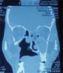
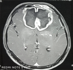
A 36-year-old male with complaints of headache, left-sided facial pain, difficulty in opening left eye for 10 days, with a history of one episode of generalized seizure that lasted for 4 min, recently detected type 2 diabetic mellitus. On generalized examination, the patient was drowsy with GCS [E2V2M5], responding to pain stimuli. Local examination revealed left-sided nasal crusts, old palatal defect, vision was bilateral greater than 3/60, and extraocular movement full. KOH was positive for mucorales. CT scan PNS revealed left-sided maxillary, ethmoidal, sphenoidal frontal sinusitis with erosion of left lamina papyreceae, left extraconal and intraconal involvement with erosion of left cribriform plate (Figure 1). MRI brain revealed additional hyperdensities noted in bilateral medial frontal, and parietal lobe with diffusion restriction (Figure 2).
Pre Op:
After discussing with the Neurosurgical team, an endoscopic trans-nasal approach to the frontal lobe abscess was preferred as frontal craniotomy is associated with higher morbidity especially taking into account the poor general condition of the patient.
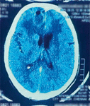
The patient underwent left-sided endoscopic sinus surgery with endoscopic endo-nasal trans-cribriform drainage of frontal lobe abscess, with retro bulbar Amphotericin B injection, with a neurosurgical team on standby in the event of unexpected complications. The mild CSF leak was managed with surgical and gelfoam sandwiched in between and fibrin glue applied with continuous lumbar drain and Acetazolamide. The eschar and pus drained from the frontal lobe abscess was sent for histopathological examination and culture and sensitivity. The patient was extubated, and the postoperative period was uneventful with improvement in the general condition gaining consciousness, and obeying oral commands. The follow-up post-operative CT brain (Figure 3) suggested of pneumatocele and minimal residual lesion, which was managed medically with Inj. Liposomal Amphotericin B, IV antibiotics, glycemic control, phenytoin. The lumbar drain was removed on third post-operative day and no CSF leak was noted.
Post Op:
Therapy with anti-fungals was continued till a cumulative dose of 2 grams was reached along with resolution of signs and symptoms of infection. IV antibiotics were added to provide coverage against bacterial infections. On the 20th post-operative day, the patient started to talk, while on the 28th post-operative day, the patient was able to walk without any support.
Discussion
This was the first case of frontal lobe abscess drainage done through an endoscopic endonasal approach successfully in our institution. In our case, open neurosurgical approach was avoided because of the poor general condition of the patient; moreover, it may lead to abscess rupture and fulminant meningitis. The minimally invasive technique of endoscopic endo-nasal trans- cribriform approach was done as the infection tract extended from the nose into the frontal lobe. The defect in the cribriform plate was identified and the frontal abscess drained per nasally.
Multiple sittings of aggressive surgical debridement often involving orbital exenteration and concurrent medical management with anti-fungal agents like Amphotericin B remains the mainstay (7, 8). In our case the frontal lobe abscess drainage was feasible due to its continuity with the cribriform plate, which made it easily accessible endoscopically without causing any complications.
Conclusion
The drainage of frontal abscesses that are in continuity with the skull base can be effortlessly performed by an endoscopic endo-nasal approach with minimal morbidity and complications. It is a minimally invasive technique that achieves effective and complete drainage of the abscess without the morbidities of a trans-cranial approach and brain retraction. The decision-making process must involve a multidisciplinary team of infectious disease specialists, neurosurgeons, and ENT surgeons. Early diagnosis and aggressive treatment can significantly curb mortality while providing better outcomes (9), with this endoscopic endonasal approach for draining frontal lobe abscesses.
References
1. Saedi B, Sadeghi M, Seilani P. Endoscopic management of rhinocerebral mucormycosis with topical and intravenous amphotericin B. J Laryngol Otol (2011) 125:807–10. doi: 10.1017/S0022215111001289
2. Chikley A, Ben-Ami R, Kontoyiannis D. Mucormycosis of central nervous system. J Fungi. (2019) 59:1–20. doi: 10.3390/jof5030059
3. Ben-Ami R, Luna M, Lewis RE, Walsh TJ, Kontoyiannis DP. A clinicopathological study of pulmonary mucormycosis in cancer patients: Extensive angioinvasion but limited inflammatory response. J Infect. (2009) 59:134–8. doi: 10.1016/j.jinf.2009.06.002
4. Bannykh SI, Hunt B, Moser F. Intra-arterial spread of Mucormycetes mediates early ischemic necrosis of brain and suggests new venues for prophylactic therapy. Neuropathology. (2018) 38:539–41. doi: 10.1111/neup.12501
5. Economides MP, Ballester LY, Kumar VA, Jiang Y, Tarrand J, Prieto V, et al. Invasive mold infections of the central nervous system in patients with hematologic cancer or stem cell transplantation (2000–2016): Uncommon, with improved survival but still deadly often. J Infect. (2017) 75:572–80. doi: 10.1016/j.jinf.2017.09.011
6. Higo T, Kobayashi T, Yamazaki S, Ando S, Gonoi W, Ishida M, et al. Cerebral embolism through hematogenous dissemination of pulmonary mucormycosis complicating relapsed leukemia. Int J Clin Exp Pathol. (2015) 8:13639–42.
7. Patron V, Orsel S, Caire F, Aubry K, Jégoux F. Transethmoidal drainage of frontal brain abscesses. Surg Innov. (2010) 17:300–5. doi: 10.1177/1553350610380933
8. Vilalta V, Munuera J. Rhino-cerebral mucormycosis: Improving the progniosis of life- threatening disease. J Infect Dis Ther. (2014) 2:1–3.


