Introduction
Otosclerosis, a main cause of conductive hearing loss, is caused by unusual bone growth around the stapes footplate, hindering its movement and sound transmission. Stapedotomy is the gold standard surgical procedure creating a small opening in the fixed stapes footplate, followed by prosthesis placement. Earlier, this procedure was performed using a microscope (MS). However, recent progress in endoscopic technology has paved the way to the exploration of endoscopic stapedotomy (ES) as a potentially less invasive technique.
ES offers several advantages over MS, such as an enhanced view of the middle ear’s intricate structures, less need to remove bone, and less disturbance of the chorda tympani nerve (1–3). These benefits could lead to improved surgical precision, lower risk, and quicker patient recovery. However, ES requires quiet a long learning curve for surgeons.
This case series, conducted in our hospital, aims to examine the safety, effectiveness, and learning process associated with ES, while also incorporating the findings from previous research in this area.
Materials and methods
This study was a case series of patients who underwent ES for otosclerosis in our hospital between January 2021 and December 2023.
Patient inclusion criteria were:
• Diagnosis of otosclerosis confirmed by audiometry and HRCT scan of the temporal bone
• Age ≥ 18 years
• Underwent primary ES under LA as the initial surgical intervention for otosclerosis.
Data collection
Patient information was extracted from both electronic health records and detailed surgical notes. Information included patient demographics, analysis of steps of ES, surgical details (e.g., type of prosthesis, use of laser or drill), audiological outcomes (pre and post AC and BC values), and any complications (intraoperative, early postoperative, and late postoperative).
Statistical analysis
Patient demographics and surgical outcomes were summarized using descriptive statistics. A paired t-test was used to analyze the differences between preoperative and postoperative air-bone gap (ABG) measurements, with a p-value of < 0.05 deemed significant.
Surgical technique
Endoscopic stapedotomy was performed under Local Anesthesia (LA) or Monitored Anesthesia Care (MAC) by a single surgeon.
Premedication and positioning
The patient was pre-medicated with IV Pethidine and Phenergan (promethazine). The patient was placed in a supine position with their head gently supported by a ring and turned toward the surgical side. The major advantage of endoscopic stapedotomy is that it does not require a rigid head position, providing flexibility for both the surgeon and the patient.
Infiltration
Local infiltration was done with 2% xylocaine and 1:200,000 adrenaline in the postauricular sulcus and external auditory canal (Four Quadrant Technique). In endoscopic surgery (ES), special attention was given to postero-superior infiltration to ensure that the infiltration sufficiently elevated and blanched the meatal flap, along with local anesthetic effect. The major advantage of ES is the close and complete visualization of infiltration in the canal wall.
(1) Tympanomeatal flap: The tympanomeatal flap in ES requires a thin flap, as in a standard stapedotomy. (For endoscopic ear surgery, a thick, wide flap in the posterior superior canal is required for bony work). Two vertical incisions—right (12−7 o’clock) then left (12−5 o’clock) and a curvilinear incision connecting both vertical incisions (at the midpoint of the annulus and mucocutaneous junction) were made. The incision should be to the depth of the periosteum, and the flap should be gradually elevated to the fibrous annulus. Care was taken to differentiate between the fibrous annulus and the chorda tympani nerve. The Posterior tympanic spine was identified, and a curved pick was used to elevate the fibrous annulus from the bony annulus. The advantage of ES in this step is the close visualization of all landmarks (fibrous annulus, chorda tympani nerve, and posterior tympanic spine) (Figure 1A). Meatal bleeding was controlled with 4% xylocaine and adrenaline-soaked cotton balls. Bipolar cautery can be supportive if needed. The instruments required were a double-ended (flag and Rosen knife), monopolar RF cautery, and a laser to reduce bleeding.

Figure 1. Tympanomeatal Flap Elevation (A), Drilling (B), Landmark Exposure (C), Removal of Stapes suprastructure (D), Footplate Perforation (E), Prosthesis Fixation (F).
(2) Tympanotomy: Middle ear tympanotomy was completed. A 4 mm endoscope is sufficient, but a 2.7 mm endoscope (0° and 30°) can be used additionally for complete evaluation of the tympanum ( meso-, pro-, hypo-, retro-, and epitympanum) and ossicular mobility, thus confirming otosclerosis. The advantages of ES in this step include close, panoramic, angled, and gentle visualization without difficulties.
(3) Otosclerosis drilling: Drilling the post-superior canal wall is required for complete visualization of the stapes footplate (oval window) (Figure 1B). The following landmarks—pyramid process, stapedial tendon, and facial nerve (horizontal segment)—must be exposed in this step (Figure 1C). It can be done with a curette or diamond burr. The diamond burr (size 1.4 mm) can be used for the posterior tympanic spine with utmost care to avoid damage to the chorda tympani nerve. Chorda tympani nerve can be repositioned for complete visualization of the oval window, superiorly and inferiorly. This step exposes the basic landmarks of Endoscopic Ear Surgery (stapes and facial nerve−horizontal segment). These landmarks should be identified before proceeding with any further steps in functional endoscopic ear surgery (FEES), Endoscopic Stapedotomy, endoscopic glomus tympanicum Excision. The incudomalleolar joint (IMJ), incudostapedial joint (ISJ), and footplate were checked for mobility. Footplate fixation must be confirmed before proceeding with stapedotomy.
(4) Prosthesis length and diameter selection: The length and diameter of the Teflon prosthesis were assessed before proceeding with stapedotomy. The diameter used in our cases is 5 mm; 4 mm can also be used, as suggested by Ugo Fisch. Length was measured using a measuring rod (4.5 mm to 5 mm) in our cases. We used Teflon prosthesis (TPP), but titanium prostheses can also be used.
(5) Stapedotomy: Stapedotomy is the major step in all stapes surgeries, requiring mastery in ear surgery. The steps can be classical or reversed, as described by Fisch et al. (4). We performed the reversed technique in cases with adequate space between the stapes crura and facial nerve. The classical technique was used in narrow spaces. The reversed technique requires more expertise than the classical. Classical steps include IS joint separation, cutting of the stapedial tendon, followed by posterior and Anterior crurotomy, Removal of the suprastructure, Footplate perforation, prosthesis insertion and fixation. Reverse steps include footplate perforation, Prosthesis insertion and fixation, IS joint separation, Cutting of the stapedial tendon, Posterior crurotomy, and Anterior crurotomy. IS joint separation can be done with a 45° angle fine pick, and the stapedial tendon can be cut with scissors (J, S, S) (Figure 1D). A diode laser (10 W) has also been tried. Posterior crurotomy was done with a 0.6 mm sized diamond burr, scissors, crurotomy and laser. Anterior crurotomy was done with similar instruments. Before proceeding to stapedotomy, the field is cleared with gelfoam and a cotton ball. The 4 mm endoscope is switched to a 2.7 mm 0° scope for visualizing the anterior crus of stapes. The advantage of a 4 mm endoscope is that it is clear, does not fog, and does not cause heat damage. For close view, 2.7 mm or 3 mm scope can be used for visualization in the tympanum, though fogging and heat damage are drawbacks.
(6) Footplate perforation and fixation: Footplate perforation is a crucial step in stapedotomy, which can be done with a perforator (3, 4, 5, 6 mm) for a 5 mm prosthesis, a laser, or a 0.6 mm diamond burr (Figure 1E). Teflon prosthesis insertion and fixation is the final step, where the prosthesis is carefully inserted into the small opening in the footplate and attached to the long process of the incus bone (Figure 1F). Prosthesis mobility was checked in both vertical and horizontal directions and the hearing outcomes of the patient were assessed on the table. ES is a single-handed technique that offers advantages in all steps and is done under local anesthesia.
(7) Sealing and final visualization: Sealing of the stapedotomy was done with ear lobule fat. This fat is very thin and offers better sealing. The endoscope 4 mm or 2.7 mm is used to visualize the footplate position, mobility, and sealing. Finally, the flap was repositioned, and the cut edges were approximated and fixed with gelfoam.
Results
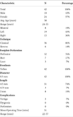
Our study included 42 individuals who met our specific criteria for endoscopic stapedotomy (ES) and underwent the procedure during the designated timeframe. The demographic and clinical details of these patients are summarized in Table 1.
In Table 2, the results of paired t-tests we used to look at the differences in hearing before and after surgery are presented. The results clearly show a substantial improvement in all three key measures of hearing air conduction (AC), bone conduction (BC), and the air–bone gap (ABG) after patients underwent endoscopic stapedotomy. They are highly statistically significant (p < 0.0001 for all).
The results demonstrate that endoscopic stapedotomy (ES) is highly effective in improving hearing function, as evidenced by the significant improvements in AC, BC, and ABG. The substantial reduction in ABG confirms the successful treatment of the conductive component of hearing loss, a hallmark of otosclerosis.
All surgeries employed Teflon prostheses with a 0.5 mm diameter. The majority of the prostheses used were 4.5 mm in length (74%), followed by 5 mm (19%) and 4.75 mm (7%). Classical techniques were predominant (86%), with the remaining 14% using reversal techniques.
The complication rates were low and comparable to those reported in the literature. Minor complications included vertigo in 7% of cases. Importantly, there were no instances of dysgeusia or tympanic membrane perforation.
The learning curve for ES was analyzed by observing the mean operating time, which was 31 min with a range of 22–57 min. A trend toward reduced operative times and fewer complications was observed with increasing surgeon experience, consistent with other studies in the field.
Discussion
Our findings support the feasibility and safety of ES in our hospital. The high success rate of ABG closure aligns with previous studies, confirming the efficacy of ES in treating otosclerosis. The low rate of major complications, particularly the absence of permanent facial nerve paralysis and sensorineural hearing loss, highlights the safety of ES in experienced hands.
The observed learning curve for ES is consistent with the findings of other studies (5, 6). As surgeons gain experience with the technique, operating time duration and complication rates tend to decrease. (This underscores the importance of specialized training and mentorship for surgeons transitioning from MS to ES).
Nikolaos et al. (7) argued that while endoscopic surgery doesn’t seem to yield significant audiological improvements compared to conventional microscopic techniques, it does result in less scutum drilling and fewer cases of postoperative dysgeusia. However, we diverge from this assessment based on our own analysis.
Contrary to Nikolaos et al. (7), Bartel et al. (8) reported that in their analysis of 573 cases, the preferred endoscope diameter was 3 mm in 51% of cases and 4 mm in 39%, while we exclusively utilize the 4mm diameter. They also found that titanium piston prostheses are used in 52% of cases, Teflon in 48%, whereas we exclusively employ Teflon prostheses. The prosthesis length was 4.5 mm and 0.6 mm diameter in 81% cases and 0.4 mm in 19%, while we use a 5 mm length with a diameter ranging from 0.4 to 0.6 mm. Furthermore, the mean surgical time reported by Bartel et al. was 55 min, whereas our average surgical time is 31 min. Results show comparably similar audiological outcomes to microscopic approaches.
Similarly, Elsamnody et al. (9) proposed that endoscopic stapedotomy presents a viable replacement for microscopic techniques, offering shorter operative times, minimal complications, and significant hearing improvement. However, we challenge their assertion that endoscopic stapedotomy is merely an alternative to microscopic surgery, as the choice between the two methods often depends on the surgeon’s expertise and preference, rather than the clear-cut superiority of one over the other. Additionally, we suggest that hearing outcomes are likely similar for both approaches.
ES has been shown to be a feasible technique in various studies, including cases of revision surgery and complex anatomy (1–3). The use of a 0° or 30°, 4 mm endoscope gives better visualization of middle ear structures, facilitating endomeatal incisions and tympanomeatal flap elevation (1–3, 5). While the surgical steps of ES mirror those of MS, adaptations are made to accommodate the use of an endoscope and one-handed techniques.
Multiple studies have reported several advantages of ES over MS in terms of a wider field of view and superior illumination compared to MS, particularly in narrow or tortuous ear canals (1–3, 5). This enhanced visualization allows for better identification and preservation of critical structures like the chorda tympani and facial nerve, potentially reducing iatrogenic injury. ES may reduce the need for substantial removal of the posterior superior canal wall, thus potentially lowering the risk of chorda tympani nerve injury and associated taste alterations (1, 2, 10, 11). Limited bone removal also potentially decreases the risk of developing iatrogenic cholesteatoma, a known complication of MS (1). Although some studies suggest potential benefits of ES in terms of faster recovery and reduced postoperative pain compared to MS, further investigation is needed (12).
ES does have its limitations: transitioning from microscope-based surgery can be difficult due to the one-handed technique and altered spatial perception (13). Precise prosthesis placement and crimping can be more difficult with ES due to limited maneuverability and the two-dimensional view offered by the endoscope. ES doesn’t require any specific endoscopic equipment or instruments, which can be universally accessible. The initial cost of acquiring endoscopic equipment may be higher than that of traditional microscopes. The proximity of the endoscope’s light source to middle ear structures raises concerns about thermal injury although studies suggest the risk is very minimal with proper technique and equipment (6).
Conclusion
Endoscopic stapedotomy is evolving as the gold standard approach for stapedotomy, comfortably performed with a single-handed technique under local anesthesia. There are no limitations in the steps of surgery in this endoscopic transcanal approach. Experience and expertise are the basic requirements for endoscopic stapedotomy. Hearing outcomes in stapedotomy depend more on the surgeon’s experience than on whether an endoscopic or microscopic technique is used.
References
1. Naik C, Nemade S. Endoscopic stapedotomy: Our view point. Eur Arch Otorhinolaryngol. (2016) 273:37–41. doi: 10.1007/s00405-014-3468-6
2. Nogueira Júnior J, Martins M, Aguiar C, Pinheiro A. Fully endoscopic stapes surgery (stapedotomy): Technique and preliminary results. Braz J Otorhinolaryngol. (2011) 77:721–7. doi: 10.1590/S1808-86942011000600008
3. Blijleven E, Willemsen K, Bleys R, Stokroos R, Wegner I, Thomeer H. Endoscopic vs. microscopic stapes surgery: An anatomical feasibility study. Front Surg. (2022) 9:1054342. doi: 10.3389/fsurg.2022.1054342
4. Fisch U, May J, Linder T. Tympanoplasty, mastoidectomy, and stapes surgery. 2nd ed. New York, NY: Thieme (2008). p. 222–53.
5. Isaacson B, Hunter J, Rivas A. Endoscopic stapes surgery. Otolaryngol Clin North Am. (2018) 51:415–28. doi: 10.1016/j.otc.2017.11.011
6. Sproat R, Yiannakis C, Iyer A. Endoscopic stapes surgery: A comparison with microscopic surgery. Otol Neurotol. (2017) 38:662–6. doi: 10.1097/MAO.0000000000001371
7. Nikolaos T, Aikaterini T, Dimitrios D, Sarantis B, John G, Eleana T, et al. Does endoscopic stapedotomy increase hearing restoration rates comparing to microscopic? A systematic review and meta-analysis. Eur Arch Otorhinolaryngol. (2018) 275:2905–13. doi: 10.1007/s00405-018-5166-2
8. Bartel R, Sanz J, Clemente I, Simonetti G, Viscacillas G, Palomino L, et al. Endoscopic stapes surgery outcomes and complication rates: A systematic review. Eur Arch Otorhinolaryngol. (2021) 278:2673–9. doi: 10.1007/s00405-020-06388-8
9. Elsamnody A, Yousef A, Taha M. Outcomes of endoscopic stapedectomy: Systematic review. Int Arch Otorhinolaryngol. (2024) 28:e165–9. doi: 10.1055/s-0043-1761171
10. Tarabichi M. Endoscopic middle ear surgery. Ann Otol Rhinol Laryngol. (1999) 108:39–46. doi: 10.1177/000348949910800106
11. Das A, Mitra S, Ghosh D, Sengupta A. Endoscopic stapedotomy: Overcoming limitations of operating microscope. Ear Nose Throat J. (2021) 100:103–9. doi: 10.1177/0145561319862216
12. Moneir W, Abd El-Fattah AM, Mahmoud E, Elshaer M. Endoscopic stapedotomy: Merits and demerits. J Otol. (2018) 13:97–100. doi: 10.1016/j.joto.2017.11.002

